Scalp Biopsy
Clinical methods for diagnosing hair loss using tissue analysis.
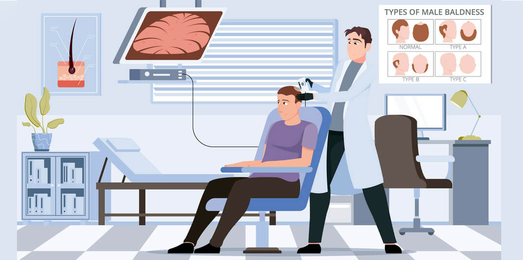
Overview of Scalp Biopsy
A scalp biopsy is a minor surgical procedure that removes a small sample of skin and hair follicles for microscopic examination. It is one of the most definitive diagnostic tools for hair loss, particularly when the cause is unclear or when a scarring alopecia is suspected.
While non-invasive methods such as trichoscopy provide valuable information, a biopsy allows direct visualization of the affected tissue, revealing changes at the cellular level that cannot be seen through other means.
When Is a Scalp Biopsy Indicated?
- Scarring alopecia is suspected: Conditions such as lichen planopilaris or frontal fibrosing alopecia require histological confirmation.
- Atypical presentation: When the clinical appearance does not match a typical pattern of hair loss.
- Failure to respond to treatment: When standard therapies have not produced expected results.
- Multifactorial hair loss: When multiple potential causes exist and need to be differentiated.
- Patient anxiety: When a definitive diagnosis provides psychological reassurance.
Biopsy Techniques
The choice of biopsy technique depends on the clinical suspicion and the information required. Each method has specific indications and advantages.
Shave Biopsy
- Description: Removes superficial layers of the skin using a scalpel or razor blade.
- Depth: Limited to the epidermis and superficial dermis.
- Indications: Useful for superficial lesions or when a full-thickness sample is not required.
- Advantages: No sutures required; minimal discomfort; quick healing.
- Limitations: Does not sample the deep dermis or subcutaneous tissue; may miss deeper pathology.
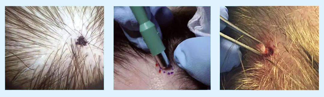
Punch Biopsy (4mm)
- Description: A circular instrument (trephine) is used to remove a full-thickness cylindrical sample of skin.
- Depth: Full-thickness to the subcutaneous fat.
- Indications: Standard for hair disorders; provides adequate tissue for both vertical and horizontal sectioning.
- Advantages: Provides a complete cross-section of the scalp; simple technique; heals with minimal scarring.
- Limitations: May require one or two sutures; minimal bleeding; small sample size may miss patchy disease.
For most hair disorders, a 4mm punch biopsy is the preferred technique. Two samples may be taken if both vertical and horizontal sectioning are planned.
Excision Biopsy
- Description: Complete surgical removal of a lesion or area of interest.
- Depth: Full-thickness, often including subcutaneous tissue.
- Indications: Used for larger lesions, suspected neoplasms, or when complete removal is therapeutic.
- Advantages: Allows complete histopathological assessment; may be curative for localized lesions.
- Limitations: Requires sutures; longer procedure; greater scarring.
Sectioning Methods
Once the biopsy sample is obtained, it is processed and prepared for microscopic examination. The way the sample is sectioned determines what information can be obtained.
Horizontal Sectioning
For hair loss evaluation, horizontal sectioning is the preferred technique. The sample is sectioned parallel to the skin surface, allowing examination of multiple hair follicles at various depths.
What horizontal sectioning reveals:
- Follicular density: The number of follicles per unit area.
- Miniaturization: The ratio of terminal to vellus-like follicles – a key feature of androgenetic alopecia.
- Anagen-to-telogen ratio: The proportion of growing to resting hairs.
- Inflammatory infiltrates: Presence of lymphocytes, plasma cells, or other inflammatory cells around follicles.
- Fibrosis (scarring): Replacement of normal tissue with fibrous tissue, indicative of cicatricial alopecia.
- Perifollicular changes: Alterations in the tissue surrounding the hair follicle.
Horizontal sectioning is considered the gold standard for diagnosing scarring alopecias and differentiating between various non-scarring conditions.
Vertical Sectioning
Vertical sectioning involves cutting the sample perpendicular to the skin surface. This provides a cross-sectional view of the skin layers.
- Useful for: Assessing the relationship between the epidermis, dermis, and subcutaneous tissue.
- Limitations: Only a small number of follicles are visible; may miss patchy inflammation.
For comprehensive evaluation, many dermatopathologists recommend both horizontal and vertical sectioning – a technique known as "transverse and vertical sectioning" – to maximize diagnostic accuracy.
What Pathologists Look For
The microscopic examination of a scalp biopsy can reveal a range of findings that guide diagnosis:
Non-Scarring Alopecias
- Androgenetic alopecia: Increased miniaturization; altered anagen-to-telogen ratio; no significant inflammation.
- Telogen effluvium: Increased telogen follicles; normal follicular density; no inflammation.
- Alopecia areata: Peribulbar lymphocytic infiltrate (a "swarm of bees" pattern); eosinophils may be present.
Scarring Alopecias
- Lichen planopilaris: Lichenoid interface dermatitis; perifollicular lymphocytic infiltrate; fibrosis.
- Frontal fibrosing alopecia: Similar to lichen planopilaris; involves the frontal hairline and eyebrows.
- Central centrifugal cicatricial alopecia: Perifollicular inflammation; fibrous tracts; loss of follicular units.
Other Findings
- Infectious causes: Fungal organisms, bacterial colonies, or parasites.
- Neoplastic conditions: Abnormal cellular growth or malignancy.
- Structural abnormalities: Abnormalities in the hair shaft or follicle architecture.
The Biopsy Procedure: What Patients Can Expect
Understanding the procedure can help alleviate patient anxiety and improve cooperation during the process.
Before the Procedure
- Informed consent: The procedure, potential risks, and expected outcomes are explained.
- Medication review: Certain medications that affect bleeding (such as aspirin or blood thinners) may be paused.
- Site marking: The biopsy site is identified and marked.
During the Procedure
- Anesthesia: Local anesthetic is injected into the scalp – a brief stinging sensation followed by numbness.
- Biopsy: A 4mm punch biopsy instrument is rotated to remove the tissue sample. This takes approximately 2-3 minutes.
- Closure: One or two simple sutures are placed. The wound is covered with a dressing.
- Duration: The entire procedure typically takes 10-15 minutes.
Patients may feel pressure but should not feel pain. Some bleeding is normal and expected.
After the Procedure
- Recovery: Minimal downtime. Patients can return to normal activities within 24 hours.
- Wound care: The dressing should be kept dry for 24-48 hours. The suture site should be kept clean.
- Suture removal: Sutures are typically removed in 7-10 days.
- Healing: The site heals completely within 2-4 weeks, with minimal scarring.
- Hair growth: Hair will grow through the biopsy site once healing is complete.
Potential Risks
- Bleeding: Minimal; pressure dressing controls it.
- Infection: Rare but possible; proper wound care minimizes this risk.
- Pain: Mild discomfort post-procedure; manageable with over-the-counter pain relief.
- Scarring: A small, often circular scar may remain; usually hidden by surrounding hair.
- Temporary hair loss: Some shedding may occur around the biopsy site; hair typically regrows.
Results and Follow-Up
Biopsy results are typically available within 7-14 days. The report is reviewed by a dermatopathologist who specializes in hair disorders.
Understanding the Report
- Clinical history: Correlates the biopsy findings with the patient's symptoms and clinical presentation.
- Histological description: A detailed description of cellular and tissue-level changes.
- Diagnosis: The specific diagnosis based on the histological findings.
- Recommendations: Suggested next steps based on the diagnosis.
Next Steps
- If a non-scarring condition is diagnosed: Medications, lifestyle modifications, or ongoing monitoring may be recommended.
- If a scarring condition is diagnosed: Anti-inflammatory medications, topical therapies, or advanced interventions may be initiated.
- If the biopsy is inconclusive: Additional tests or repeat biopsy may be considered.
- Psychological support: A definitive diagnosis often reduces anxiety and provides clarity.
Key Clinical Points
- Scalp biopsy with horizontal sectioning is the standard for diagnosing scarring alopecias.
- Scalp biopsy is safe, well-tolerated, and provides definitive diagnostic information.
Making the Decision
A scalp biopsy is a valuable diagnostic tool when the cause of hair loss is uncertain. It provides definitive information that guides treatment decisions and helps patients understand their condition. While the procedure may sound intimidating, it is quick, relatively painless, and generally well-tolerated. With appropriate clinical follow-up, the benefits of an accurate diagnosis typically outweigh the minor risks.